What are Bow-legs?
Bowlegs are a deformity around the knee in which the leg has deviated inwards with respect to the thigh. When a child with bowlegs stands with his or her feet together and toes pointed straight ahead, the knees do not touch. The medical term for bowlegs is “genu varum” (Figure 1).

What is Normal (Physiological) Bow-legs And Knock-knees?
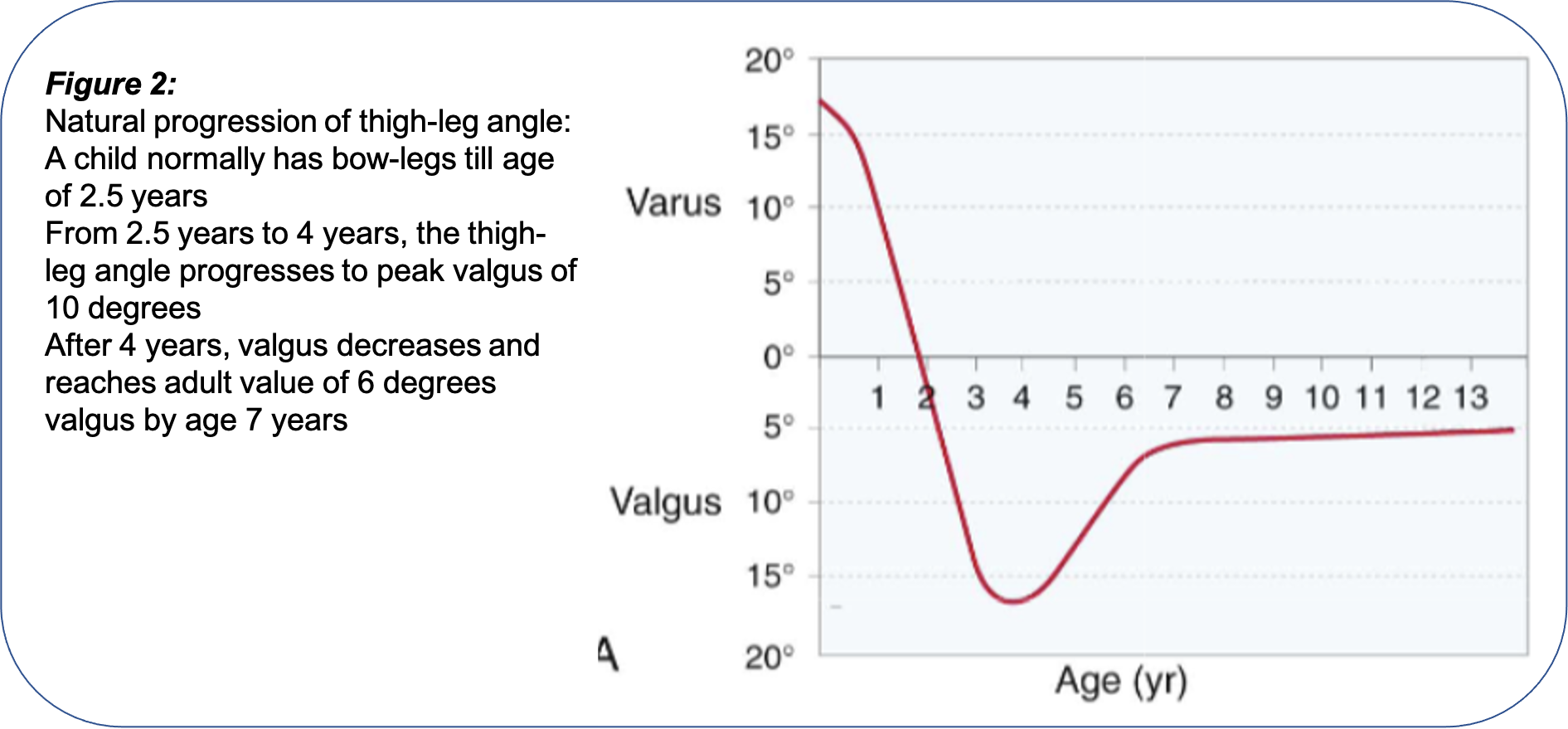
In a child, the angle between the thigh and leg (called the “thigh-leg angle”) follows a certain pattern and keeps changing with age.
- A newborn child normally has bowlegs, and the thigh-leg angle is between 10 to 15 degrees varus. This angle gradually decreases in the second year of life and the leg becomes straight by 2.5 years ago. This normal varus angle which persists till the age of 2.5 years is called “Physiological bowlegs” (Figure 2).
- After the age of 2.5 years, the child normally develops knock-knees and the thigh-leg angle reaches a peak valgus angle of about 10 degrees by the age of 4 years. This normal valgus angle is called “Physiological knock-knees” (Figure 2).
- After the age of 4 years, the thigh-leg angle again decreases and reached an adult angle of about 6 degrees valgus by the age of 8 years (Figure 2).

So, What Are The Red-Flag Signs To Identify Abnormal Or Pathological Deformities Which Need Treatment?
Any deformity which fails to follow the above normal pattern is deemed to be abnormal.
- Deformity persisting beyond the abovementioned normal age range. E.g Bowlegs in a 7 years old child (Figure 3).

- The deformity is more severe than the above-mentioned angles. E.g: Bowlegs angulation of 20 degrees in a 2 years old child (Figure 4).

- Asymmetrical deformity (Figure 5).

- Deformity in association with an underlying skeletal dysplasia (Figure 6).

- Deformity occurring following an infection/trauma (Figure 7).

- Deformity occurring due to underlying metabolic bone disease (Figure 8).

How is a child with bowlegs or knock-knees clinically assessed?
Clinical assessment of a child with bowlegs or knock-knees consists of:
- Assessment of severity of the deformity
- Differentiation of physiological bowlegs from Blount’s disease
- Assessment of location of the deformity
- Assessment of progression of the deformity
- Assessment of severity of deformity: This is done by measurement of thigh-leg angle. The thigh-leg angle is measured as the angle joining ASIS-centre of patella and center of patella-center of the ankle. This angle is measured with the knee extended and patella facing forwards (Figure 9).

- Assessment of location of deformity: Bowlegs or knock-knees deformity can occur due to deformity either in the thigh bone (femur) or in the leg bone (tibia). Whether the femur or tibia is deformed can be assessed by the knee flexion test. In this test, the deformity in the leg is assessed with the knee extended (straight) and knee flexed (folded). If the deformity disappears after knee flexion, the deformity lies in the femur (thigh bone). On the other hand, if the deformity persists even after knee flexion, the deformity lies in the tibia (Figures 10 and 11).


- Differentiation between Physiological bowlegs and Blount’s disease: This is done by performing the “Cover Test”. In this test, the orientation of the knee is assessed after covering the distal third of the tibia with a pillow.
In the case of physiological bowlegs, the deformity is located in the distal leg and so after covering the distal leg, the orientation of the knee as assessed by thigh leg angle is in valgus (Figure 12).

On the other hand, in a child with Blount’s disease, the deformity is in the proximal leg and so the varus deformity at the knee remains visible even after the distal leg is covered in the cover test (Figure 13).

- Assessment of progression of deformity: Assessment of worsening or improvement of deformity is done by measuring the inter-malleolar distance (in case of knock-knees), or, inter-condylar distance (in case of bowlegs). If the distance is increasing, it indicates worsening of the deformity. On the other hand, if the distance is decreasing it indicates improvement of deformity (Figures 14 and 15).


What is The Treatment Of Bowlegs And Knockknees?
The first step in the treatment of bowlegs and knock-knees is establishing the diagnosis, which is done on the basis of clinical examination, blood investigations (blood levels of Calcium, Vitamin D, etc.), X-rays and in special situations advanced imaging like MRI, CT-scan, etc.
In most cases, bowlegs/ knock-knees are normal (physiological) in which case parents need to be reassured that the deformity will automatically resolve with age.
In cases where the deformity is abnormal (pathological), treatment may consist of:
- Medical treatment: Supplementation with Calcium and Vitamin D is needed in cases where the deformity is secondary to a nutritional deficiency of these nutrients. After correction of the blood levels of Calcium and Vitamin D, deformities usually resolve gradually over a period of 1.5 to 2 years
- Surgical treatment: is needed in most cases of pathological (abnormal) bowlegs and knock-knees. Surgical treatment is broad of two types:
-Growth modulation (guided growth) with eight plates: The eight plate is a metallic device that is implanted in the growing bone straddling the growth plate. The eight plate functions by guiding the bone growth in such a way that the deformity corrects with growth. This technique is an ideal modality for younger children with healthy growth plates with sufficient growth remaining (Figure 16).

Corrective Osteotomy: Osteotomy is a surgical procedure in which the deformed bone is cut at the level of deformity, the deformity is corrected and the bone is fixed in the corrected position with implants to maintain the correction. It is a larger surgical undertaking than a growth plate and needs to be performed in older children or in children with damaged growth plates in whom the potential for remaining growth is limited (Figure 17).

–Posted in the public interest by the Division of Children’s Orthopaedics, Pinnacle Orthocentre Hospital. For more information, mail drsvvaidya@gmail.com. To schedule an appointment, call 7028859555.


0 Comments