Congenital Muscular Torticollis Treatment: What Parents Need to Know
Case 1
Clinical presentation:
A 7 years old male child presented with neck deformity noted by parents since birth, restriction of neck movements and facial asymmetry. He had undergone physiotherapy when he was one year old, however the deformity failed to resolve.
On examination, the child’s neck is tilted to the Left. The sternocleidomastoid muscle on the Left side of the neck was taut. The child’s cervical spine movements were restricted (Figure 1).
X-ray of the Cervical spine did not reveal any bony problem.
In view of these findings, diagnosis of Left Congenital Muscular Torticollis was confirmed.

Figure 1: Left Congenital muscular torticollis. Note the neck tilt to Left and tight sternocleidomastoid muscle (red arrow)
Congenital Muscular Torticollis
The child was treated by Left sternocleidomastoid unipolar release surgery in which the tight muscle is lengthened. In the post-operative period, the child underwent physiotherapy in the form of muscle stretching and range of motion exercises.
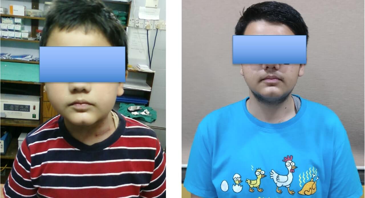
The neck deformity was corrected at 2 months after surgery. The correction was maintained at 5 years following surgery (Figure 2).

Figure 2: Post-operative correction of torticollis deformity. The correction is well-maintained at 5 years follow-up.
Case 2
Clinical presentation:
A 5 years old male child presented with neck deformity since birth. He had a taut Left sternocleidomastoid muscle. X-ray Cervical spine did not reveal any bony abnormality, thereby establishing the diagnosis of Left Congenital Muscular Torticollis.
He underwent surgery for Left sternocleidomastoid muscle release surgery which resulted in correction of deformity in the post-operative period.

Figure 3: Left Congenital Muscular Torticollis in a 5 years old male child corrected after surgery for Left sternocleidomastoid muscle release surgery.
Discussion:
What’s Congenital Muscular Torticollis?
It’s a condition wherein a muscle in the neck called the Sternocleidomastoid is tight. As a result, the neck is tilted to the side of the tight muscle and movements of the neck are restricted.
In long-standing uncorrected cases, facial asymmetry may arise with the face being flattened on the side of torticollis.
What causes CMT?
While it was earlier thought that CMT occurs due to injury at the time of birth, recent reports have debunked this theory and we now know that the Stermocleidomastoid is tight since birth, cause for which is not yet understood.
How is CMT diagnosed?
CMT is diagnosed clinically by identification of the tight muscle on the side of deformity. However your doctor may order Xrays to be sure that there is no underlying bony deformity.
How is CMT treated?
If CMT is detected early, it can be treated by Physiotherapy. Physiotherapy in CMT is mainly aimed at stretching the tight SCM muscle. Physiotherapy is successful in correcting deformity in approximately 50% cases.
Children who fail physiotherapy need surgery.
When is surgery needed?
Surgery is usually needed if the CMT deformity fails to resolve with physiotherapy by the age of one year. However in that case, surgery is withheld till the age of 3 years
What is actually done in the surgery?
Surgery in CMT is involves release of the tight Sternocleidomastoid muscle either at the lower end only (just above the collar bone, called Unipolar release) or both at lower and upper ends (behind the ear, called Bipolar release) in case of severe deformities.
What is the post-operative protocol?
Postoperatively, the child needs to undergo physiotherapy after one week. The torticollis deformity usually completely corrects by 6 weeks after surgery. Facial asymmtery if present prior to surgery takes about 1 to 1.5 years for correction.
What about the scar?
The surgery is done through a cosmetic incision, so the scar is hardly visible after surgery.
_ By Dr Sandeep Vaidya, Paedictric Orthopaedic Surgeon, Pinnacle Orthocentre Hospital. For more information, mail drsvvaidya@gmail.com or Call on: +91 7028859555.
