Introduction:
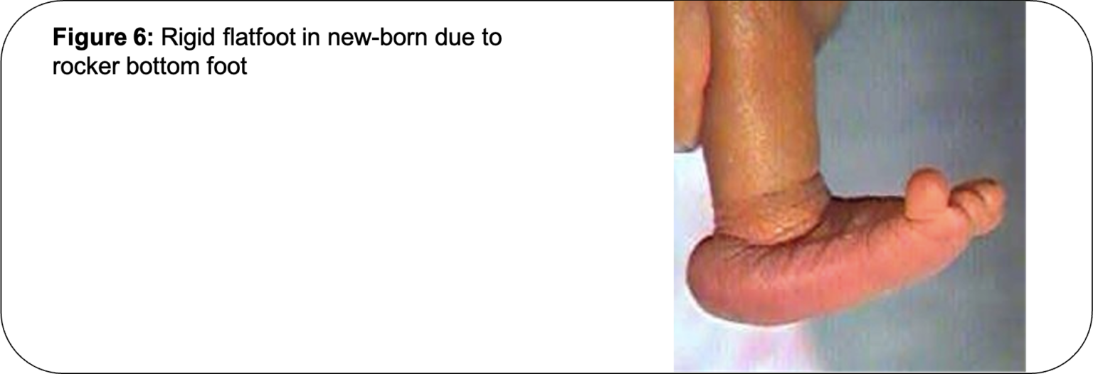
Congenital Vertical Talus (CVT) is also called Rocker Bottom Foot. It is a birth deformity of the foot.
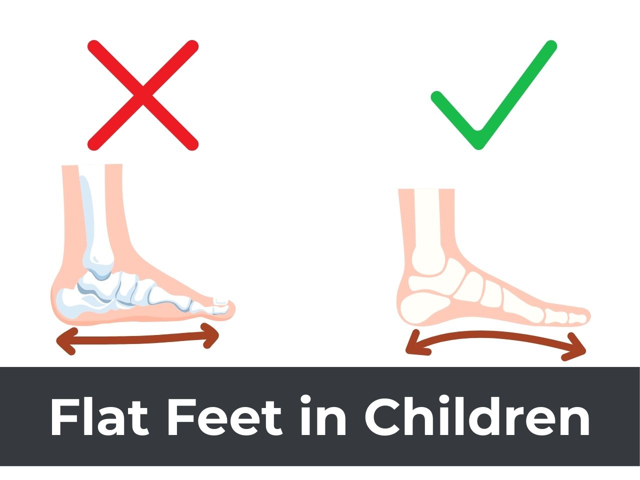
Normally, the sole of the human foot has an arch and is concave when viewed from the side (Figure 1).

In Congenital Vertical Talus, the sole of the newborn child’s foot is convex when viewed from the side (Figure 2).

Normal bony anatomy of the foot:
The talus is the bone in the ankle joint, that lies between the shinbone (tibia) above and the heel bone (calcaneum) below. The rest of the bones of the foot lie in front of it. The normal orientation of the talus bone is in a horizontal direction (Figure 3).

Bony anatomy in CVT:
Congenital Vertical Talus is a birth deformity in which the talus bone, instead of being oriented in a horizontal direction, is directed vertically. There is dislocation of the talo-navicular joint (Figure 4).

Associated conditions:
A child may have CVT as an isolated deformity or as part of a collection of multiple deformities like Arthrogryposis Multiplex Congenital (AMC) or Meningo-myelocele (MMC) or certain syndromes. Hence, a thorough complete evaluation of the child is necessary to rule out these associated conditions.
Congenital Vertical Talus Surgery Treatment
Minimally invasive treatment:
Initial treatment in a new-born child with CVT consists of non-operative treatment. This involves the application of serial plaster casts to the child’s leg to stretch out the deformity. After application of serial plaster casts, if the satisfactory alignment of the talo-navicular joint is obtained, a minimally invasive surgery consisting of the release of the tight Achilles tendon (heel cord) and open pinning of the talo-navicular joint is performed. This minimally invasive treatment protocol is called the “Dobbs’ ” or “Reverse Ponseti’s” protocol. This minimally invasive treatment protocol may not be successful in all cases, but nevertheless, serial plaster casts should be pursued prior to open surgical treatment in all cases because it helps to stretch out the tight soft tissues and makes surgical treatment less extensive and technically easier.
Surgical treatment:
Open surgical treatment is performed in CVT if deformity fails to correct with the minimally invasive treatment protocol. A significant percentage of children with CVT will need open surgery. Surgery consists of the circumferential release of the sub-talar joint, and, lengthening of certain tight tendons and ligaments in the foot. This allows the talus to become mobile following which its orientation is corrected from the vertical to a horizontal position. The talus is then held in the correct position by inserting a metal wire (called K-wire) across the talo-navicular joint (Figure 5).

After surgery, a plaster cast is applied. The K-wire and cast are removed after 4 to 6 weeks.
A brace is usually prescribed to maintain the correction. Your doctor will recommend you to follow up regularly for a few years to monitor the growth of the foot and to check for recurrence of the deformity (Figure 6).

this article is contributed by Dr. Sandeep Vaidya, Paediatric Orthopaedic Surgeon, Pinnacle Orthocentre Hospital, Thane. Dr. Vaidya is also available for consultations at BJ Wadia Children’s Hospital, Mumbai; Ajit Scan Centre, Kalyan; and Ace Children’s Hospital, Dombivli. For more information, call 7028859555/ 8879970811/ (022)25419000/ 25429000 OR email drsvvaidya@gmail.com.


0 Comments