Introduction:
Osteoid osteoma is a relatively uncommon but benign bone tumor that can cause significant pain and discomfort. While it is generally not life-threatening, the symptoms it produces can greatly impact a patient’s quality of life. In this article, we will explore what osteoid osteoma is, its symptoms, diagnosis, and available treatment options.
What is Osteoid Osteoma?
Osteoid osteoma is a small, non-cancerous (benign) tumor that forms in the bone. It usually develops in the long bones, such as the femur (thigh bone) or tibia (shin bone), but can occur in any bone. This tumor is composed of osteoid tissue, which is an immature bone.
Symptoms of Osteoid Osteoma:
The hallmark symptom of osteoid osteoma is pain, which is often worse at night and typically responds to over-the-counter pain medications like aspirin or ibuprofen. The pain is caused by the release of prostaglandins, substances that induce inflammation.
Other common symptoms include swelling and limited joint movement near the affected area.
Diagnosis of of Osteoid Osteoma:
Diagnosing osteoid osteoma can be challenging, as its symptoms can mimic other musculoskeletal conditions. Imaging studies such as X-rays, CT scans, or MRIs are commonly used to identify the tumor and assess its size and location (Figure 1).
In some cases, a bone scan may be recommended to help pinpoint the exact location of the tumor.

Figure 1: Osteoid osteoma of the Right femur neck. In this case, the tumour was not visualised on X-ray. CT scan revealed the diagnosis of osteoid osteoma.
Osteoid Osteoma Treatment:
Several treatment options are available for osteoid osteoma, and the choice of treatment depends on the tumor’s size, location, and the severity of symptoms.
Non-Surgical Approaches:
Pain Medication: Over-the-counter nonsteroidal anti-inflammatory drugs (NSAIDs) like ibuprofen or aspirin can provide temporary relief from pain.
However, the pain returns after the effect of the medicine wears off. Hence this is only a temporary measure till surgery if performed.
Minimally Invasive Surgery (CT scan guided techniques):
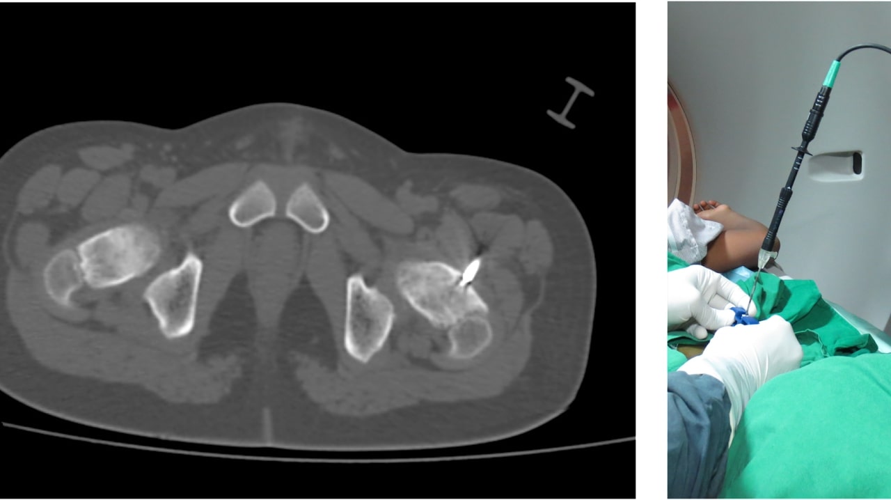
- CT scan guided Radiofrequency Ablation (RFA): This procedure involves using a special needle, under CT scan guidance, to deliver heat to the tumor, destroying it while minimizing damage to surrounding healthy tissue (Figure 2).
- CT scan guided drilling: This procedure involves drilling of the osteoid osteoma tumour under CT scan guidance.
Open surgery: In some cases, particularly if non-surgical approaches are not effective or if the tumour location is not suitable for minimally invasive surgery, open surgical removal of the tumor may be necessary.

Figure 2: Treatment of the case in Figure 1 with CT guided RFA ablation. Under CT scan guidance, the tip of the Radio Frequency Ablation (RFA) probe is inserted into the centre of the osteoid osteoma. The tip of the probe is heated to 100 degrees Celcius which results in destruction of tumour tissue. Patient experiences almost immediate pain relief after the surgery.
Prognosis:
The prognosis for osteoid osteoma is generally excellent. Most patients experience relief from pain after appropriate treatment, and the tumor is not known to metastasize or spread to other parts of the body.
Conclusion:
Osteoid osteoma may present challenges, but with proper diagnosis and treatment, patients can regain control over their lives. If you or someone you know is experiencing persistent bone pain, it’s essential to consult with a healthcare professional.
Early detection and intervention can make a significant difference in managing symptoms and preventing complications associated with osteoid osteoma.
– By Dr Sandeep Vaidya, Paedictric Orthopaedic Surgeon, Pinnacle Orthocentre Hospital.
For more information, mail drsvvaidya@gmail.com/ call 7028859555.


0 Comments