Brief Clinical Case Summary:
A 14-years-old female child presented with progressive bilateral genu valgum (knock knees), which was affecting her gait and causing discomfort.
The deformity had become more pronounced over the last two years, and she was facing functional difficulties, particularly while walking and running.
She was diagnosed with bilateral genu valgum due to idiopathic causes. (Figure 1).

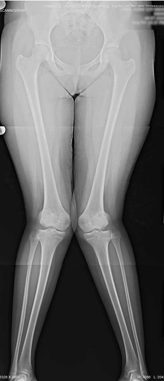
Figure 1: Clinical photograph and pre-operative X-ray of 14 years old female child with bilateral genu valgum deformity
What was done?: Hemi-epiphysiodesis versus Osteotomy
- The girl was post-menarchal
- The growth plates around the knee and triradiate cartilage were closed.
It was therefore concluded that the girl didn’t have growth remaining, and therefore her deformity could not be corrected by hemi-epiphysiodesis technique.
In view of this, the girl underwent a corrective osteotomy surgery. In this case, we performed a distal femur medial closed wedge osteotomy fixed with plate and screws.
Since the magnitude of deformity correction was more than 20 degrees, prophylactic common peroneal nerve decompression was performed at the same sitting (Figure 2).
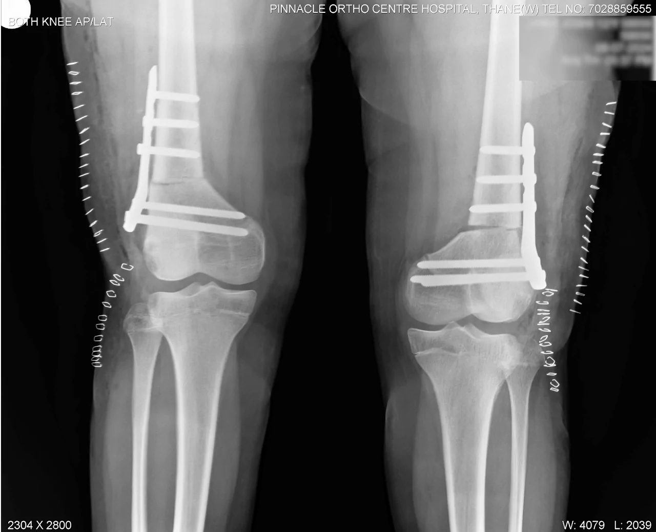
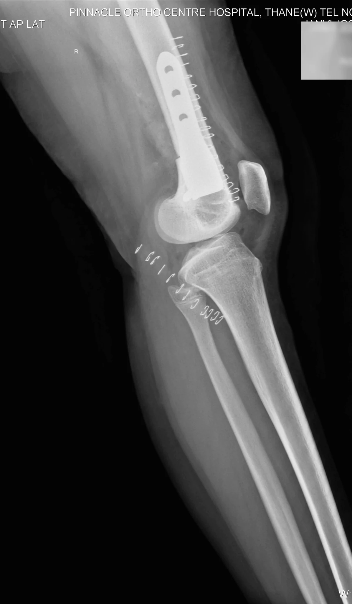
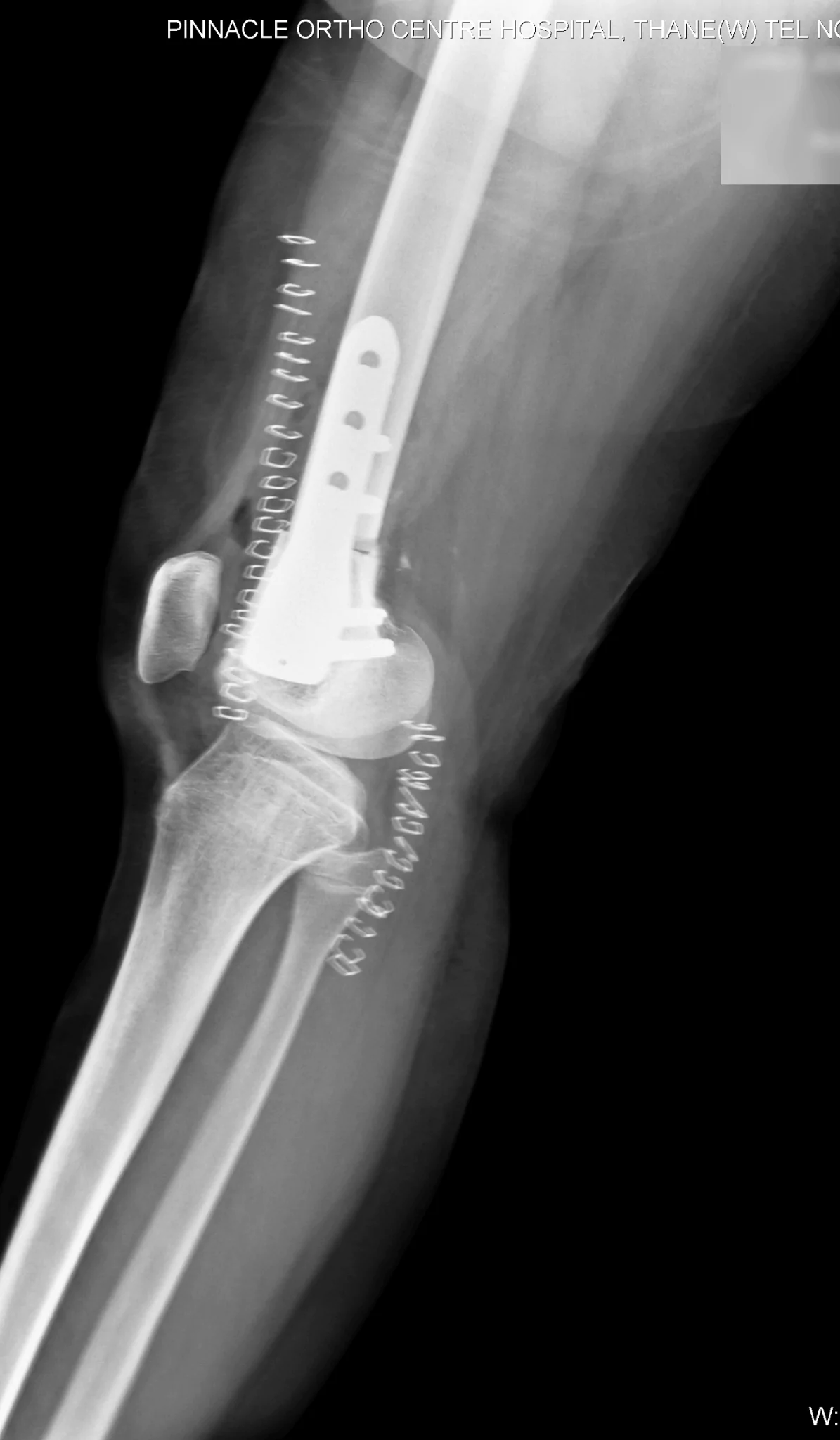
Figure 2: Post-operative X-ray bilateral distal femur medial closed wedge osteotomy
Post-operative correction of deformity:
After surgery, the child’s deformity was corrected. Clinically, the thigh leg angle and inter-malleolar distance was normalised. Radiologically, the aLDFA was normalised.
The osteotomy healed over a period of 6 weeks following which the child was permitted full weight bearing mobilisation (Figure 3).

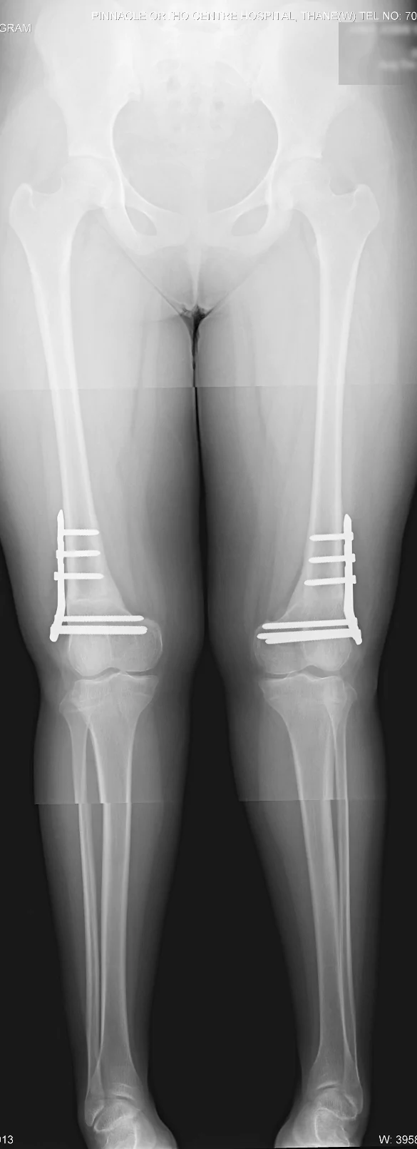
Figure 3: 1 year post-operative clinical photograph and X-ray shows correction of deformity
Conclusion:
Osteotomy is an effective method of correction of genu valgum deformity. It is done in older children after growth has ceased.
_ By Dr Sandeep Vaidya, Paedictric Orthopaedic Surgeon, Pinnacle Orthocentre Hospital.
For more information, mail drsvvaidya@gmail.com/ call 7028859555.



0 Comments