We report the management of a 45-year-old man with a ring stuck in his dominant thumb for 3 weeks causing vascular and neural compromise and deep-seated infection. Previous reports have described rings embedded in the digits, though we were not able to find any reports of such rings causing ischaemia.
We performed staged reconstruction to salvage the thumb. The thumb viability was confirmed after ring removal and the infection was treated as per culture sensitivity report. Both digital nerves were reconstructed with sural nerve graft and the circumferential soft tissue defect was covered with bilobed first dorsal metacarpal artery flap from index and middle finger. At the 6-month follow-up, the patient had useful function of the thumb with well-settled donor site on index and middle finger.
Level of Evidence: Level V (Therapeutic)
Introduction
A ring is the most common ornament in many cus- toms and religions. It is traditionally worn as a symbol of love or as decorative jewellery.
However, rings are not without risk. Tight rings can result in swollen digits and must be removed immediately before they threaten digit viability. Persistent constriction by the ring can result in pressure necrosis of skin, loss of tissue, infection and compromised vascularity of the digit. The usual method of treatment is to simply cut the ring and remove it from the finger. Occasionally patients with psychological illness or addictions may refuse to remove the stuck ring.
We report a patient with known psychological illness with a delayed presentation of a swollen digit due to a stuck ring and discuss the staged management of patients who present late with a stuck ring.
Case Report
A 45-year-old man presented with a ring embedded in his dominant right thumb. He gave history of sustain- ing some blunt trauma to the right thumb 3 weeks ear- lier and subsequent swelling of the thumb followed by a wound and foul-smelling discharge.
He also noticed that the thumb was numb and cold to touch and he had difficulty moving the interphalangeal joint. He gave his- tory of treatment for depression. On examination, the right thumb was swollen distal to the embedded ring at the level of mid-shaft of the proximal phalanx shaft with delayed capillary filling (5 seconds) and hypoaesthesia on both the radial and ulnar aspects of pulp.
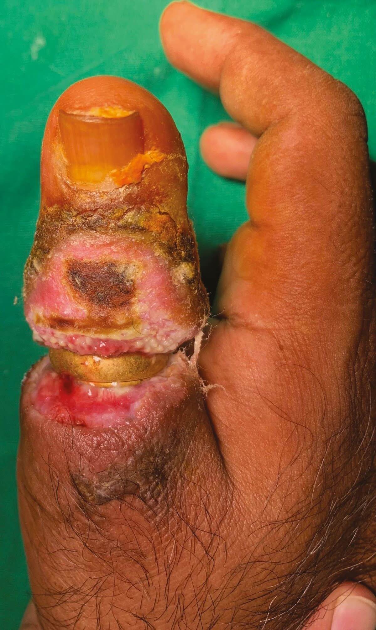
The ring was embedded into the soft tissue around the proximal pha- lanx circumferentially (Fig. 1). A foul-smelling discharge was noted. The active and passive flexion and extension of the interphalangeal joints were less than 10º.
The active range of motion of the first metacarpophalangeal joint was 15º extension to 60º flexion. On percussion at palmar region, he had positive localised Tinel sign. There was no evidence of chronic osteomyelitis or fracture on X-ray and the ring was visible at mid shaft level of proxi- mal phalanx of thumb.
After 7 days of regular wound care, the wound showed healthy granulation tissue. We confirmed the viability of thumb. The capillary filling status was the same as before surgery. Therefore, we did not opt for any vascu- lar intervention at this stage. Soft tissue cover by local and distant pedicle flaps were discussed with the patient.
We planned the second stage of surgery to reconstruct the digital nerves and provide soft tissue cover.
The patient was counselled for surgery under regional anaesthesia. The ring was cut with a special ring cutter. Bruner incision was made on the palmar aspect after removal of the ring. The neurovascular bundles on the ulnar and radial side were explored.
Both digital vessels were noted to be thrombosed. Both digital nerves were noted to be fibrotic. We noticed chronic inflammation surrounding the tendons of the flexor and extensor policies longus, though the tendons were in continuity.
The per- iosteum was breached, though the bone was intact and there were no signs of infection in the bone. All unhealthy and infected soft-tissue was debrided. The tissue was sent for gram staining, culture and sensitivity.
The wound was left open after debridement and dressed with a moist gauze and a thumb spica slab applied after surgery. The capillary filling remained the same after surgery.
The culture grew a mixed growth including Staphylococcus aureus and coagulase-negative staphylococci, which were sensitive to gentamicin and clindamycin.
Antibiotics were given under guidance of infectious disease specialist.
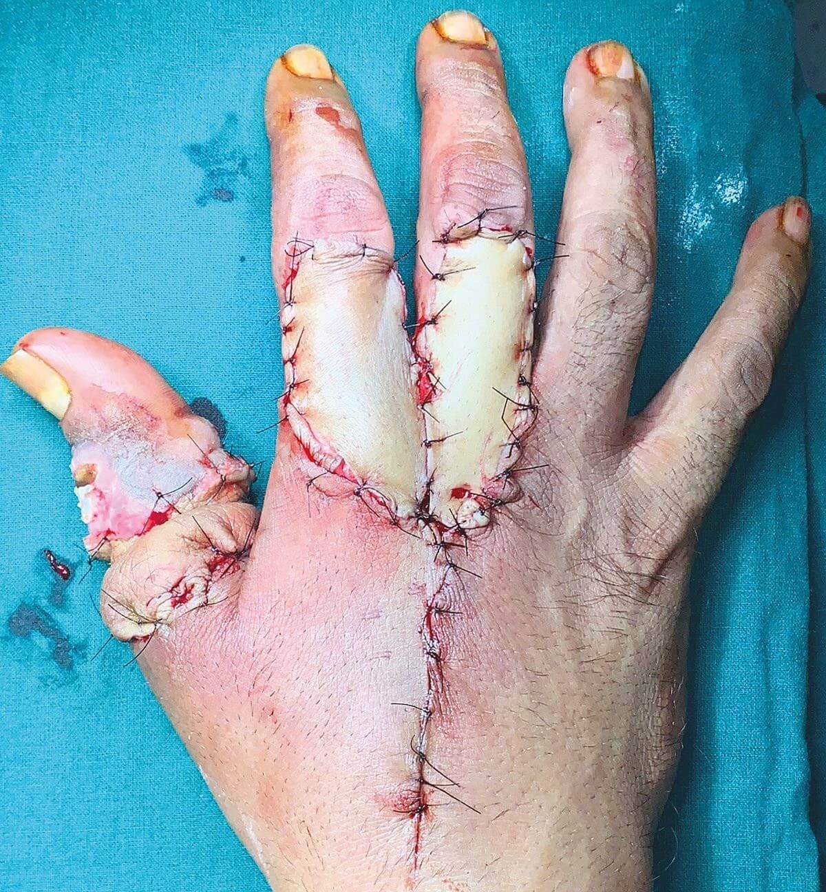
Fig. 3. Circumferential defect on thumb
The digital nerve reconstruction (both radial and ulnar) was done using a sural nerve graft using single cable after excis- ing neuroma (Fig. 2).
The patient had a circumferential defect of the thumb with a width of 1.2 cm exposing the reconstructed digital nerves and the extensor and flexor policies longus. A local flap was chosen as the patient was obese and under psychiatric treatment.
A bilobed flap based on the first dorsal metacarpal artery (FDMA) was used to cover the defect and the flap donor defect covered with a full thickness skin graft harvested from ulnar aspect of forearm (Fig. 3). After surgery, the thumb was viable with a capillary refill time of 5 seconds.
The wounds were dressed and thumb immobilised in a back slab for 2 weeks. The flap settled well in 6 weeks and donor site on index and middle finger took 8 weeks to heal completely.
At his final follow-up at 6 months, he had 20º of flexion at the interphalangeal joint of thumb. His two-point dis- crimination on the radial and ulnar aspect of the thumb were 8mm and 12 mm, respectively.
The capillary refill time improved to 3 seconds. The opposition, abduction and rest of the hand function was satisfactory as shown in Figures 4 and 5.
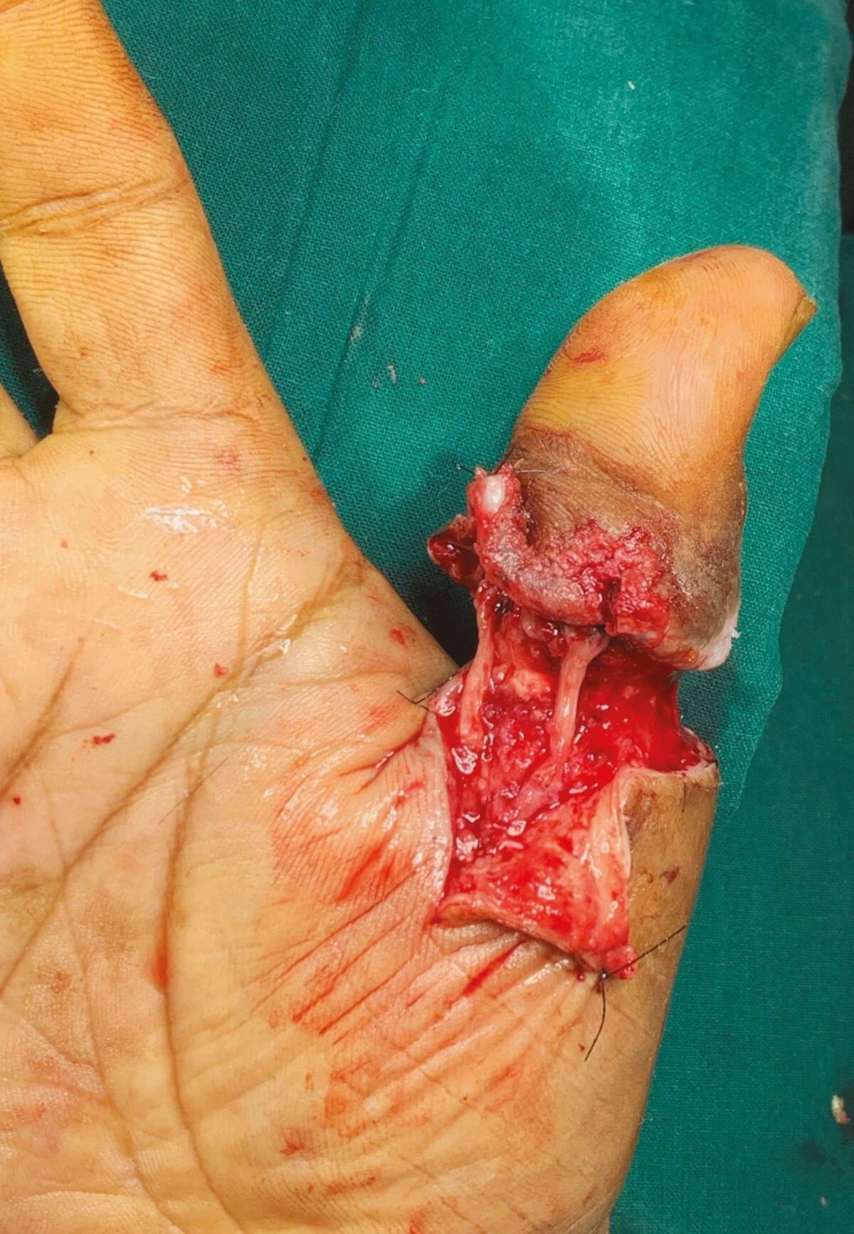
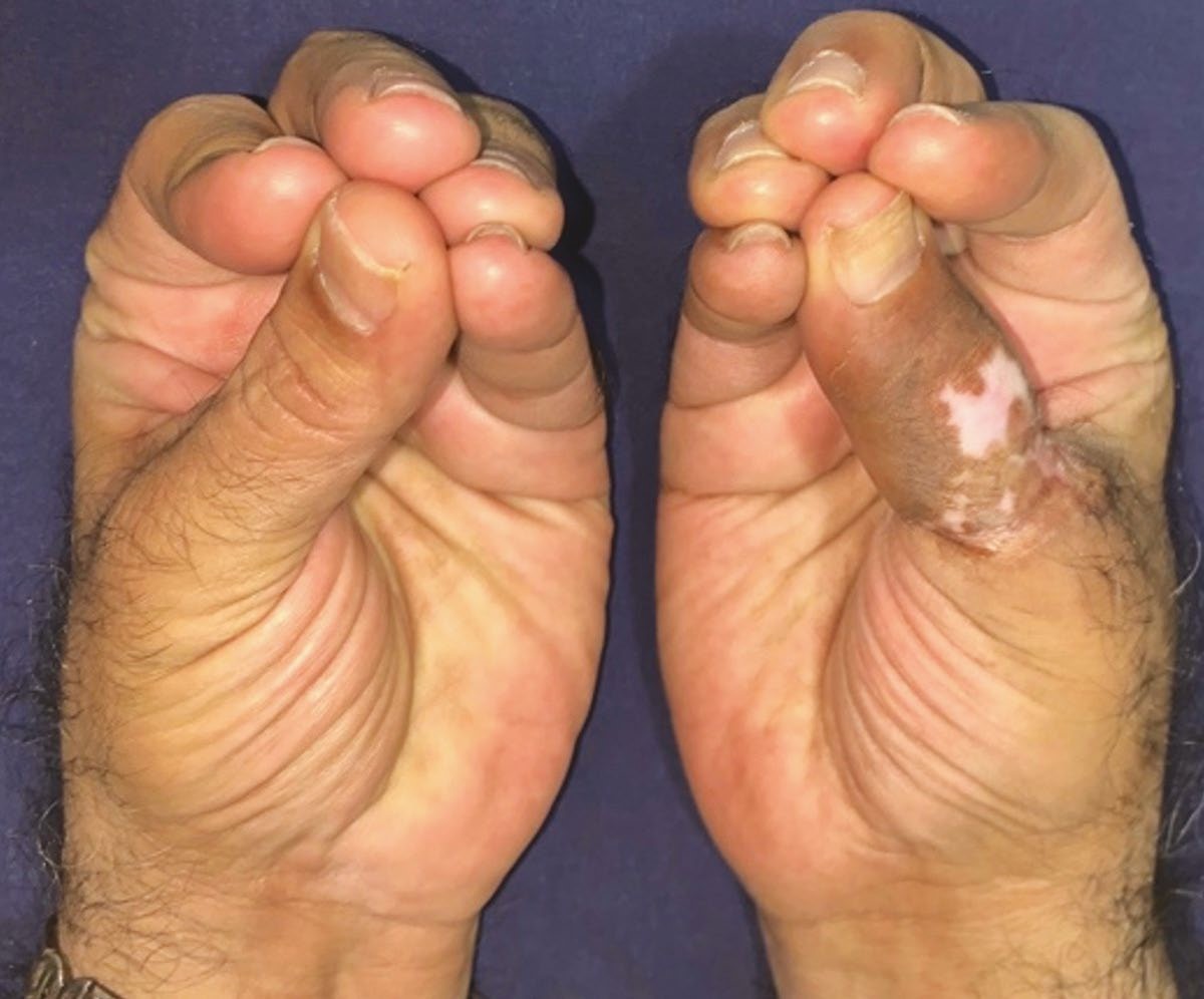
Fig. 4. Opposition of thumb at 6 months
Discussion
Many people choose to wear a ring or ‘wedding band’, which is usually forged from metal. Commonly, ring entrapment can occur secondary to the swelling of a digit.
The swelling in digit can be a result of trauma or other medical conditions. It is possible to get the ring stuck in digit secondary to pregnancy, allergic reaction or infec- tion, or simply because of a tight-fitting ring.
Ring acts as a tourniquet and leads to digital swell- ing. This will cause disruption to the lymphatic drainage exacerbating digital oedema. Progression of the swelling. Able to use a shorter piece of string because their tech- nique did not cover the entire finger.
There are various flaps described to cover the defect of thumb.9 Bilobed second dorsal metacarpal artery-based island flap described by Xu Zhang et al is appropriate for circumferential defects at proximal phalanx level.10 In chronic presentations of a stuck ring, the involvement of each anatomical tissue apart from skin need to be ana- lysed properly to plan proper reconstruction to save the digit and provide good function.
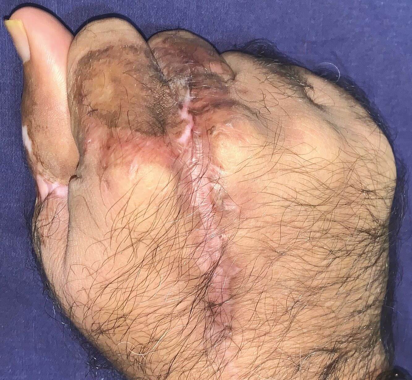
Fig. 5. Metacarpophalan- geal joint function
will then cause disruption to the venous drainage lead- ing to venous congestion. Eventually chronic compres- sion causes soft tissue erosion with arterial and neural ischaemia as well.
There are well-established methods for entrapped ring removal in the emergency department such as the winding technique, which uses thread or rub- ber band to compress the finger or using manual ring cut- ters to saw the ring.1 Rarely, a ring can become embedded into the soft tissue of a digit to the extent that it causes infection, vascular compression, neural compression and tendon adhesions leading to stiff non-functional digit.
This unusual phenomenon has previously been described as ‘embedded ring syndrome’.2 It is described mainly in digits apart from thumb.
Most reports of embedded ring syndrome describe acute management that includes ring removal, local wound debridement, primary wound closure or regu- lar dressing as a management protocol.
Rings can also result in complex traumatic avulsion injuries to the finger when the ring is caught on an object and forcefully pulled. These injuries can necessitate a range of treatments, from simple wound closure through to microvascular repair and amputation.
3 Removal of rings from swollen fingers has been discussed by several authors who have explained the process by which a piece of string can be used to pull the ring by putting the thread around the finger dis- tal to the ring, covering the finger proximal to the ring and at last pulling off the ring by unwinding the string.
4–6 Mizrahi and Lunski,7 and Belliappa,8 also used string but they wound the string proximal to distal.
Declarations
Conflict of Interest: The authors do NOT have any potential conflicts of interest with respect to this manuscript.
Funding: The authors received NO financial support for the preparation, research, authorship and/or publication of this manuscript.
Ethical Approval: Ethical approval not required for case reports.
Informed Consent: The patient was informed that this case study would be submitted for publication, and he provided a written consent.
There is no information (names, initials, hospital identification numbers or photographs) in the sub- mitted manuscript that can be used to identify patients.
Acknowledgements: None.
References
1. Kalkan A, Kose O, Tas M, Meric G. Review of tech- niques for the removal of trapped rings on fingers with a proposed new algorithm. Am J Emerg Med. November, 2013;31(11):1605–1611. https://doi.org/10.1016/j.ajem.20 13.06.009.
2. Fraser KE, Jamison DA. Embedded-ring syndrome. Ann Emerg Med. June, 1995;25(6):856–857. https://doi.org/10
.1016/s0196-0644(95)70225-3.
3. Bamba R, Malhotra G, Bueno RA Jr. Ring avulsion inju- ries: A systematic review. Hand (N Y). January, 2018;13(1): 15–22. https://doi.org/10.1177/1558944717692094.
4. Frary T. A few brief tips: Ring removal. J Am Acad Phys Assist. 1990;3:156.
5. Cresap CR. Removal of a hardened steel ring from an extremely swollen finger. Am J Emerg Med. May, 1995;13(3):318–320. https://doi.org/10.1016/0735-6757 (95)90210-4.
6. Leider M. Removal of a ring from a swollen finger with- out injuring the ring. J Dermatol Surg Oncol. April, 1980;6(4):298–299. https://doi.org/10.1111/j.1524-4725.1 980.tb00863.x.
7. Mizrahi S, Lunski I. A simplified method for ring removal from an edematous finger. Am J Surg. March, 1986;151(3):412–413. https://doi.org/10.1016/0002-9610 (86)90480-0.
8. Belliappa PP. A technique for removal of a tight ring. J Hand Surg Br. February, 1989;14(1):127. https://doi.org/ 10.1016/0266-7681(89)90035-1.
9. Rehim SA, Chung KC. Local flaps of the hand. Hand Clin. May, 2014;30(2):137–151. https://doi.org/10.1016/j.hcl.20 13.12.004.
10. Zhang X, Yang L, Shao X. Use of a bilobed second dorsal metacarpal artery-based island flap for thumb replantation. J Hand Surg Am. June, 2011;36(6):998–1006. https://doi. org/10.1016/j.jhsa.2011.03.006.



0 Comments