Brief Clinical Case Summary:
Ms. ABC, a 8-year-old child, presented to our clinic with a significant cubitus varus deformity of the right arm. The deformity, commonly referred to as a “gunstock deformity,” developed following a supracondylar fracture of the humerus sustained approximately 6 years prior.
The initial treatment involved conservative management with cast application. However, despite this intervention, the fracture healed in a malunited position, leading to a noticeable deformity.
On clinical examination, the child exhibited a humerus-ulna angle of 25 degrees, hyperextension of 20 degrees, and hypersupination of 30 degrees, all components of the cubitus varus deformity (Fig-1 ).
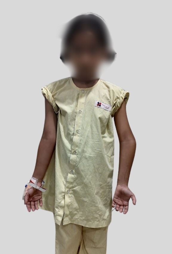
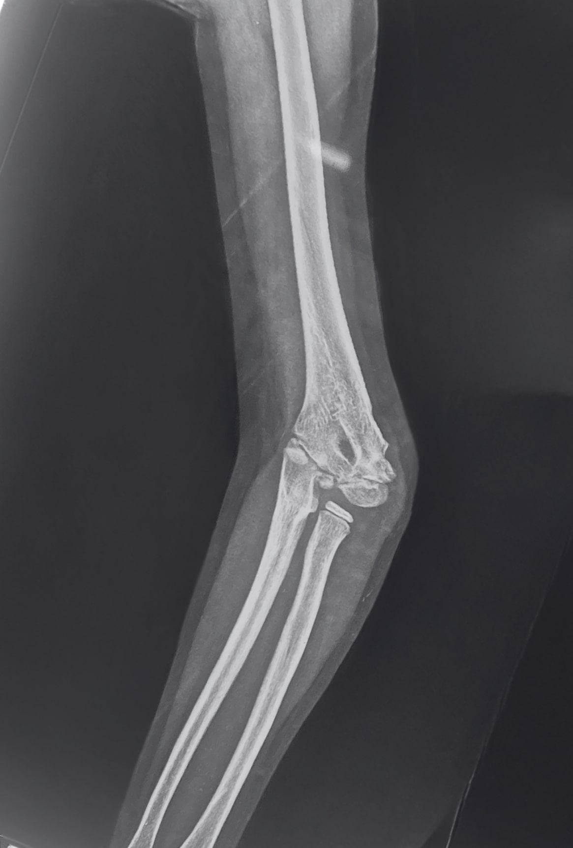
Figure 1: Clinical photograph and pre-operative X-ray of 8 years old child with cubitus varus deformity
The cubitus varus had both functional and aesthetic implications. The parents reported concerns about the child’s appearance and potential future limitations in activities involving the right arm. Given the significant degree of deformity and the impact on the child’s quality of life, surgical correction was deemed necessary.
Brief description of surgery done:
The patient underwent a right distal humerus lateral oblique closing wedge osteotomy. This procedure was chosen due to its efficacy in correcting angular deformities while preserving limb length.
The surgery was conducted under general anesthesia, with careful preoperative planning to determine the exact degree of correction needed.
Intra-operatively, a lateral approach was utilized to access the distal humerus. A closing wedge osteotomy was performed, carefully excising a wedge of bone to allow for realignment.
The osteotomy was angled obliquely to provide a broader bone surface for healing and to reduce the risk of lateral prominence post-surgery.
Intra-operative fluoroscopy was used throughout the procedure to ensure the accuracy of the correction. This imaging modality allowed for real-time visualization of the bone alignment and confirmed that the desired correction was achieved. The osteotomy was then stabilized using K-wires, which provided adequate fixation while minimizing the risk of complications.
Post-operative imaging confirmed the successful correction of the deformity, with the humerus-ulna angle restored to a more physiological position (Figure 2 and 3).
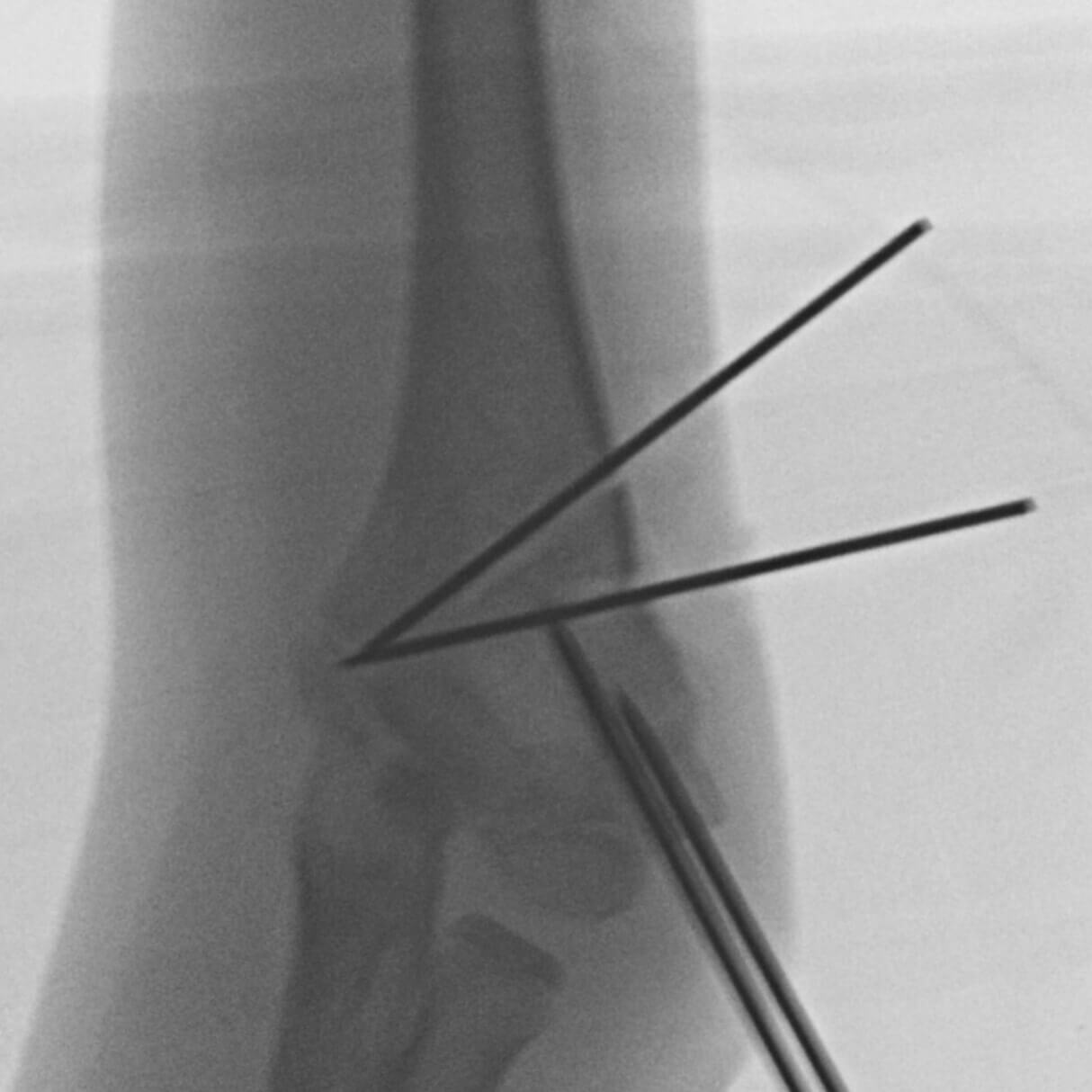
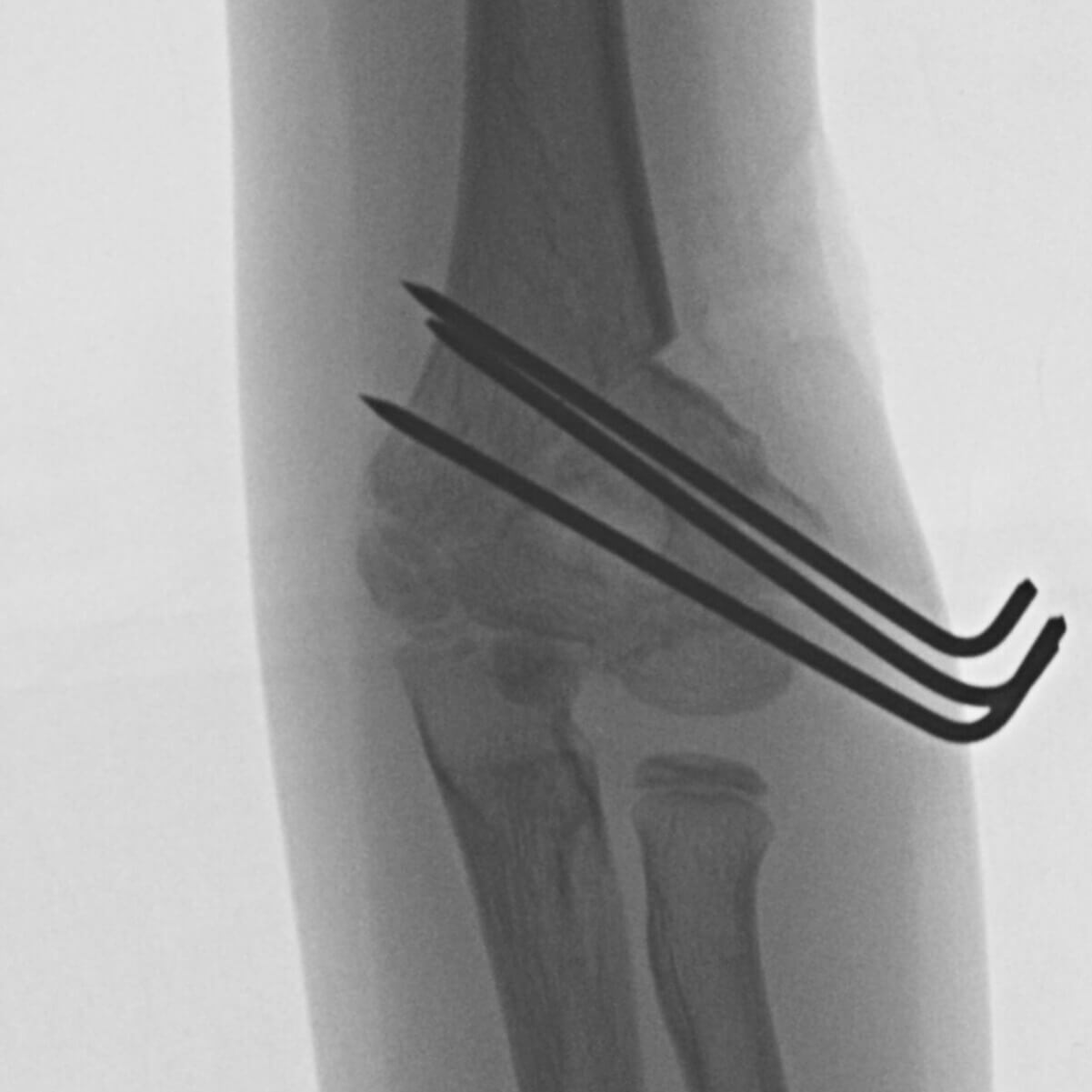
Figure 2: Intra-operative C-arm images
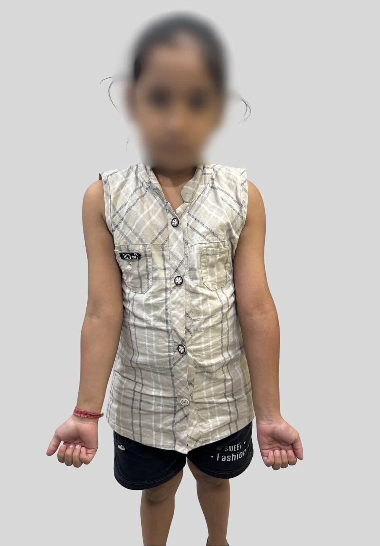
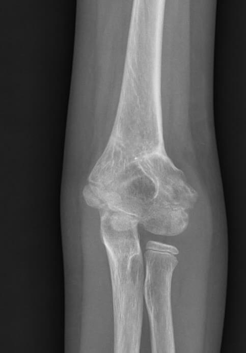
Figure 3: Followup post-operative clinical photograph and X-ray
Brief Overview of the Ailment and Available Treatment Options:
Cubitus varus, a common sequela of pediatric supracondylar humerus fractures, is characterized by a varus (inward) angulation at the elbow. This condition often arises from malunion of the fracture, particularly when the initial fracture was displaced or poorly reduced.
The deformity not only affects the cosmetic appearance of the arm but can also impair elbow function, leading to limitations in activities that require full range of motion or strength.
Treatment for cubitus varus varies depending on the severity of the deformity and the presence of any functional impairments. In mild cases, observation may be sufficient, especially if the deformity does not interfere with daily activities or is not a cosmetic concern.
However, in moderate to severe cases, surgical intervention is often necessary to correct the alignment and restore normal function.
Surgical options include various osteotomy techniques, with the lateral closing wedge osteotomy being a preferred method due to its effectiveness in correcting the deformity while maintaining the integrity of the growth plate and overall limb length.
This procedure involves the removal of a bone wedge from the lateral side of the distal humerus, allowing for realignment of the bone segments. Fixation methods, such as K-wires or plates, are used to stabilize the osteotomy site during the healing process.
In the presented case, the decision to proceed with a lateral closing wedge osteotomy was based on the degree of deformity and the functional impairment observed.
The procedure successfully corrected the cubitus varus, with the patient expected to achieve full functional recovery and a significant improvement in the cosmetic appearance of the arm.
This case underscores the importance of early and accurate intervention in cases of supracondylar fractures to prevent complications like cubitus varus. It also highlights the effectiveness of surgical correction for managing significant deformities, ensuring both functional and aesthetic outcomes for the patient.
This successful correction of cubitus varus demonstrates our commitment to providing high-quality pediatric orthopedic care.
_ By Dr Sandeep Vaidya, Paedictric Orthopaedic Surgeon, Pinnacle Orthocentre Hospital. For more information, mail drsvvaidya@gmail.com/ call 7028859555.


0 Comments